

1) Verify data readiness and labeling quality
Confirm that DICOM images, metadata, and study structure are retained end-to-end so downstream models can interpret anatomy and acquisition context. Create an inventory ai medical imaging of modalities, scanners, and protocols, because variation in reconstruction kernels and slice thickness can materially affect model performance. If your dataset is fragmented across vendors or departments, plan for normalization steps and a clear data governance workflow.
Next, validate labeling practices with a checklist for ground truth. Determine whether labels come from radiologist consensus, structured reports, or curated annotations, and document inter-reader agreement where possible. Ensure that images and labels are aligned correctly at the study, series, and slice level, with checks for off-by-one mistakes and mismatched laterality. Finally, define exclusion criteria for poor-quality scans, incomplete series, or studies with missing metadata so your training and evaluation remain trustworthy.
2) Build workflow fit with clinical and operational checks
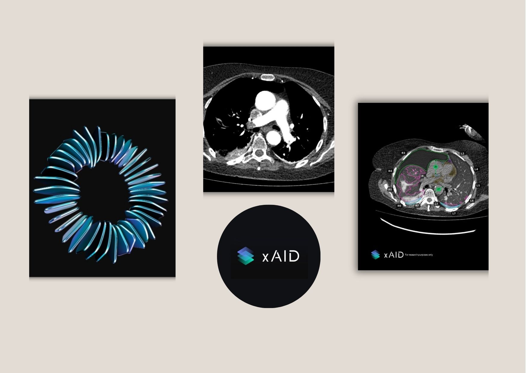
AI in radiology succeeds when it integrates into the existing reporting rhythm rather than interrupting it. Map your end-to-end workflow from patient scheduling to image acquisition, triage, and final dictation, then identify where decision support adds the most value. Use a checklist to define the intended output: heatmaps ai in radiology for region localization, priority flags for time-sensitive findings, automated measurements, or structured drafting of report sections. For each use case, specify who reviews the output, how often it triggers, and what actions the radiologist should take when the system is uncertain.
Operational readiness matters as much as model accuracy, so include checks for throughput, queuing, and fail-safe behavior. Define service-level expectations for latency, such as how quickly results should appear after images are ingested. Establish fallback logic when inputs are incomplete or when the model confidence is low, so the radiologist can proceed without delays. Also prepare training and change-management materials that explain interpretability limits, escalation paths, and how to document AI-assisted decision-making in your local quality system.
3) Ensure safety, compliance, and measurable performance
Use a compliance checklist to cover privacy, security, and regulatory expectations for diagnostic software. Validate encryption in transit and at rest, role-based access controls, audit logs, and strict data retention policies that fit your clinical setting. Confirm that your deployment model aligns with privacy requirements, including whether processing occurs on-premises, in a managed environment, or through a hybrid approach. If you handle outpatient imaging, ensure your governance supports consistent patient data handling and traceability across studies.
Then measure performance with clinically meaningful metrics rather than relying on generic scores. Include sensitivity and specificity for the targeted tasks, calibration for probability estimates, and subgroup analysis across patient demographics and scanner types. Conduct reader studies or retrospective validations that compare AI-assisted reporting against baseline workflow performance, capturing both accuracy and time-to-report. Finally, track real-world drift by monitoring input distribution changes, new acquisition protocols, and drift in measurement outputs, then define a re-validation cadence tied to your internal quality indicators.
Conclusion
A practical checklist approach reduces risk by turning adoption into a sequence of verifiable steps: data quality, workflow fit, and measurable safety. When each item is documented and tested, teams can move from pilots to stable clinical integration with clearer accountability and fewer surprises. This is especially important when AI is intended to support radiologists in producing faster, more consistent interpretations without replacing clinical judgment. For outpatient imaging centres and teleradiology providers aiming to streamline head, chest, and abdomen CT reporting, xaid.ai focuses on diagnostic efficiency designed to support accurate radiology workflows. The goal is to help teams integrate intelligent technology into reporting processes while maintaining confidence in performance and operational reliability.
